Cerebral Cortex of Developing Human Brain Mapped Using High-Quality MRI Scan Data

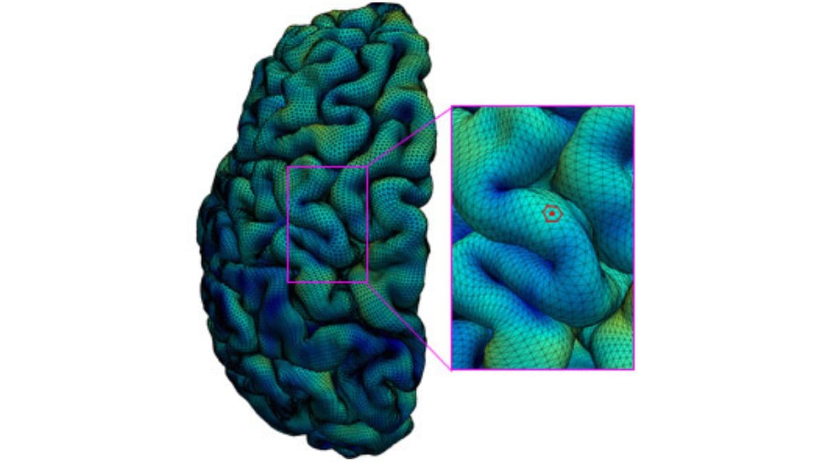
Scientists have successfully mapped the surface of the cerebral cortex of the young human brain in impressively high resolution. The mapping, done using high-quality magnetic resonance imaging (MRI) data, showed development in the key regions of the brain from two months before an individual’s birth to two months after it. Researchers believe that this achievement will likely aid future research on brain development and may serve as a new approach for studying brain development conditions like autism and schizophrenia. Cerebral cortex is a sheet of brain cells that wraps around the brain and is the most evolutionary and advanced region of the brain. It is larger in humans than any other mammal and is responsible for functions like language abilities, and abstract reasoning.

From the third trimester of pregnancy to the first two years of life, dynamic cortical development is witnessed. The cortex tends to get thick in this period and grows rapidly in terms of surface area by forming complex cortical folds. Researchers have linked disruptions in this cortical thickening and expansion to schizophrenia and autism. But, due to the lack of high-resolution mapping of this period, scientists have failed to gain a deeper understanding of this developmental stage in the fetal-to-toddler age range.

In the new study, researchers from the University of North Carolina Health Care collected a set of 1,037 high-quality MRIs of infants from the third trimester to the two years age interval. The scan data was then analysed by the team using computer-based image processing methods. With this, they divided the cortical surface into a virtual mesh having tiny circular areas and measured the surface expansion rate for each of these areas.

The team was able to define 18 distinct regions and found that they correlated well with their existing knowledge of the cortex’s functional organs. “All these regions show dramatic expansion in surface area during this developmental window, with each region having a distinct trajectory,” said Gang Li, PhD, associate professor of radiology at the UNC School of Medicine. Li is the senior author of the study published in Proceedings of the National Academy of Sciences.

The map revealed that each region of the cortex had the same developmental path as its counterpart in the opposite hemisphere. In addition, the team could also spot sex differences in the development. As per Li, the mapping has given a new insight into the brain development.

Now, the team is aiming to cast the net wider and use the approach to study scan datasets of children with autism or other neurodevelopmental conditions.

For the latest tech news and reviews, follow Gadgets 360 on Twitter, Facebook, and Google News. For the latest videos on gadgets and tech, subscribe to our YouTube channel.

Facebook to Roll Out Major Updates to Horizon Worlds; More Details at Connect Event: Mark Zuckerberg

Xiaomi NoteBook Pro 120G, Smart TV X Series to Launch in India on August 30: All Details


Check out our Latest News and Follow us at Facebook

Original Source

AI Algorithm That Recognises Anomalies in Brain May Help Treat Epilepsy

A multinational research team led by UCL has developed an artificial intelligence (AI) programme that can identify minute brain anomalies that lead to epileptic seizures. The algorithm used in the Multicentre Epilepsy Lesion Detection project (MELD), reports the locations of abnormalities in cases of drug-resistant focal cortical dysplasia (FCD), a major cause of epilepsy, was developed using more than 1,000 patient MRI scans from 22 international epilepsy centres.

Brain regions known as FCDs have evolved improperly and frequently lead to drug-resistant epilepsy. Surgery is usually used to treat it, however, finding the lesions on an MRI is a constant problem for doctors because MRI scans for FCDs can appear normal.

The scientists employed about 300,000 places throughout the brain to quantify cortical properties from the MRI scans, such as how thick or folded the cortex/brain surface was.

The system was then trained on cases that experienced radiologists had classified as either having FCD or being a healthy brain based on their patterns and attributes.

In general, the algorithm was successful in identifying the FCD in 67 percent of instances in the cohort, according to the results, which were published in Brain (538 participants).

Radiologists had previously been unable to uncover the abnormalities in 178 of the patients based on their MRI results; however, the MELD algorithm was able to detect the FCD in 63 percent of these cases.

This is crucial because, if doctors can identify the anomaly in the brain scan, surgery to remove it could result in a recovery.

Mathilde Ripart, a co-first author from the UCL Great Ormond Street Institute of Child Health, stated: “We focused on developing an AI system that was interpretable and could assist physicians in making decisions. A crucial step in that process was demonstrating to the doctors how the MELD algorithm generated its forecasts.

Dr Konrad Wagstyl, a co-senior author from the UCL Queen Square Institute of Neurology, added: “This algorithm might make it easier to identify these concealed lesions in epileptic children and adults, which would increase the number of patients who could potentially benefit from brain surgery to treat their condition and enhance cognitive function. In England, epilepsy surgery could help about 440 kids a year.”

Epilepsy is a severe neurological illness that affects 1 percent of the world’s population and is marked by recurrent seizures.

About 600,000 people in the UK are impacted. The majority of epilepsy patients can be treated with pharmaceuticals, although 20-30 percent of them do not benefit from them.

FCD is the most frequent cause in children who have had surgery to treat their epilepsy, and it is the third most frequent cause in adults.

Furthermore, FCD is the most frequent reason for epilepsy in people who have a brain anomaly that cannot be seen on an MRI scan.

Dr Hannah Spitzer, a co-first author from Helmholtz Munich, stated: “Our system automatically learns to detect lesions from thousands of patient MRI scans. It is capable of accurately identifying lesions of various sorts, forms, and sizes, including several that radiologists had previously overlooked.

Dr Sophie Adler, a co-senior author from the University College London’s Great Ormond Street Institute of Child Health, added: “We believe that this technology may assist to discover abnormalities that are now being missed that cause epilepsy. In the long run, it might make it possible for more epilepsy patients to undergo possibly curative brain surgery.

This FCD detection study makes use of the biggest MRI cohort of FCDs to date, making it capable of identifying all FCD subtypes.

Study limitations

The 22 hospitals involved in the study used various MRI scanners from around the world, making the algorithm more robust but also perhaps affecting its sensitivity and specificity.


Check out our Latest News and Follow us at Facebook

Original Source
New Study Explains How Human Brain Developed Differently Than Neanderthals

Experiments on mice have helped scientists identify some key differences in the development of brains in modern humans and our closest relative, Neanderthals. After our ancestors split from the Neanderthals, some one hundred amino acids underwent changes and spread to almost all modern humans. The reason behind this change had puzzled scientists all this while. However, six of the amino acid changes were found to have occurred in the three proteins that are instrumental in distributing chromosomes or the carriers of genetic material to the daughter cells during cell division in our body.

To dig deeper into the cause, researchers from the Max Planck Institute of Molecular Cell Biology and Genetics in Germany and the Max Planck Institute for Evolutionary Anthropology have introduced modern human variants in mice. The six amino acid positions are the same in both mice and Neanderthals.

Hence, the mice provided the scientists with a model to study human brain development. “We found that three modern human amino acids in two of the proteins cause a longer metaphase, a phase where chromosomes are prepared for cell division, and this results in fewer errors when the chromosomes are distributed to the daughter cells of the neural stem cells, just like in modern humans,” explained geneticist Felipe Mora-Bermúdez. He is the lead author of the study published in Science Advances.

Investigating if the set of amino acids in Neanderthals had the opposite effect, researchers introduced ancestral amino acids in brain organoids. These are small organ-like structures that can be grown in cell culture dishes in the lab using human stem cells. Brain organoids mimic the aspects of early human brain development.

In this case, the team noted that the metaphase was shorter while the number of errors in chromosome distribution was also more. Mora-Bermúdez described that the amino acids in modern humans were responsible for fewer mistakes in chromosome distribution. “Having mistakes in the number of chromosomes is usually not a good idea for cells, as can be seen in disorders like trisomies and cancer,” said Mora-Bermudez.


For the latest tech news and reviews, follow Gadgets 360 on Twitter, Facebook, and Google News. For the latest videos on gadgets and tech, subscribe to our YouTube channel.

Samsung Galaxy Z Flip 4 Colour, Integrated Storage Options Spotted on Official Site


Check out our Latest News and Follow us at Facebook

Original Source

Scientists Discover Human Brain Is More Hotter Than Previously Thought

Our human brain performs a variety of activities and functions throughout the day. The temperature of human brain keeps changing throughout out the day. Now, according to new research, the normal temperature of the human brain swings significantly more than previously assumed, and this could be an indication of good brain function. The study states that some areas of the deep brain can reach temperatures of up to 40 degrees Celsius, albeit this varies depending on sex, time of day, and other factors. When compared to this, the average oral temperature in humans is normally less than 37 degrees Celsius. Researchers believe that this isn’t an indication of malfunction, but rather evidence that the brain is functioning normally. Data from brain-injured patients in intensive care, where direct brain monitoring is commonly required, has previously been used in human brain temperature studies.

Using a brain scanning tool called magnetic resonance spectroscopy, researchers were recently able to detect brain temperature in healthy persons (MRS). However, till now, MRS had not been utilised to investigate how brain temperature varies during the day, or how one’s ‘body clock’ influences this.

The new study is the first to build a 4D map of healthy human brain temperature. By displaying how much brain temperature changes by brain area, age, gender and time of day, this chart disproves a number of previously held ideas.

The study also looked at data from traumatic brain injury patients, finding that the presence of daily brain temperature cycles are highly linked to survival. These findings may aid in the diagnosis, prognosis and therapy of brain injuries.

The results of the study were published in the journal Brain.

The report states that “to interpret patient analyses”, the researchers “recruited 40 healthy adults (20 males, 20 females, 20–40 years) for brain thermometry using magnetic resonance spectroscopy”. On a single day, participants were scanned in the morning, afternoon, and late evening.

In healthy participants, brain temperature ranged from 36.1 degrees Celsius to 40.9 degrees Celsius, states the report.

Female brains were 0.4 degrees Celsius warmer on average than male brains. Because most females were scanned in the post-ovulation phase of their cycle, their brain temperature was roughly 0.4 degrees Celsius warmer than females measured in the pre-ovulation phase. This difference was most likely caused by the menstrual cycle.

Dr John O’Neill, Group Leader at the MRC Laboratory for Molecular Biology, told UK Research and Innovation that the most striking discovery is that the healthy human brain can achieve temperatures that would be diagnosed as fever elsewhere in the body. In the past, such high temperatures have been measured in persons with brain injuries, but they were considered to be caused by the injury. The researchers discovered that the temperature of the brain dips before people go to sleep at night and rises during the day. There’s evidence to suspect that daily variance is linked to long-term brain health, which is something the researchers like to look at next.

The researchers are now hoping that the 4D brain temperature map can be used as a reference guide for what a healthy brain should look like. However, much more data from a far bigger group of people is needed to make it truly useful.


Check out our Latest News and Follow us at Facebook

Original Source

Exit mobile version